Spectral-domain optical coherence tomography has quietly transformed my CEA caseload over the past six years. What was once a subjective judgement call in ambiguous dogs is now a layered cross-sectional scan showing exactly which retinal and choroidal strata are affected. OCT does not replace ophthalmoscopy, but it has changed how I interpret borderline findings and how I counsel breeders with equivocal results.
How OCT Works in the Canine Eye
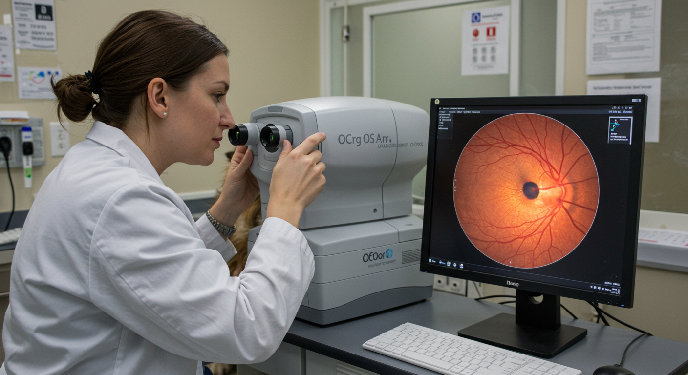
Optical coherence tomography uses a low-coherence light source, most commonly around 840 nanometres for spectral-domain systems, to produce cross-sectional images of ocular tissue at near-histologic resolution. The instrument splits the source light between a reference arm and a sample arm. When reflected light from the retina is recombined with the reference beam, the resulting interference pattern is decomposed by Fourier analysis into an A-scan showing tissue reflectivity at each depth.
Stacking hundreds of A-scans across a two-millimetre line produces a B-scan, the familiar cross-sectional OCT image that clinicians interpret. Modern spectral-domain OCT achieves axial resolution of approximately 5 micrometres, which is fine enough to distinguish individual retinal layers and the choroidal architecture underneath. For CEA, this resolution is transformative because the pathology — choroidal hypoplasia — occurs precisely in the tissue layers OCT visualizes best.
What the Scan Shows in a Normal Canine Fundus
In a healthy dog's retina, OCT reveals a predictable laminar architecture. The inner retinal layers appear as alternating bright and dark bands corresponding to the nerve fibre layer, ganglion cell layer, inner and outer plexiform layers, and nuclear layers. Below these lies the photoreceptor inner and outer segment junction, then the retinal pigment epithelium as a bright hyperreflective line, and finally the choroid beneath with its characteristic vascular pattern visible as alternating dark lumens and bright stromal tissue.
A healthy choroid has consistent thickness across the retina, typically 140 to 200 micrometres in the central fundus of medium-to-large breed dogs. The choriocapillaris, the innermost vascular layer, shows uniform hyperreflectivity on OCT. This normal anatomy is the reference against which all CEA findings are interpreted.
Choroidal Hypoplasia on OCT
In CEA-affected dogs with choroidal hypoplasia, OCT reveals exactly why the ophthalmoscopic view looks pale. The choroid in the affected region is markedly thinner than normal, sometimes measuring less than 50 micrometres. The normal vascular pattern is disorganized. The retinal pigment epithelium overlying the hypoplastic choroid may appear attenuated or absent. These findings are consistent across breeds and correlate with the visible fundus lesion.
The clinical value of OCT in choroidal hypoplasia is not diagnostic — an experienced ACVO diplomate can identify classic choroidal hypoplasia on ophthalmoscopy alone. The value is quantitative. OCT allows me to measure choroidal thickness in microns, photograph and archive the cross-section, and track any subtle changes across annual examinations. This is particularly valuable for the borderline cases discussed in our grading overview.
Colobomas on OCT
Colobomas are the higher-severity CEA finding where tissue is not merely thin but frankly absent. OCT reveals colobomas as dramatic excavations extending into or through the choroid and sclera. The surrounding retina is often displaced around the coloboma margin, and OCT can clearly show whether the retina remains attached at the edges of the defect or whether subclinical traction is present.
This last point is clinically important. Colobomas predispose to retinal detachment, and OCT can sometimes detect incipient traction before a full detachment develops. Dogs with large colobomas benefit from annual OCT surveillance regardless of whether vision has changed. The detachment-surveillance rationale is covered in detail in the retinal detachment emergency guide.
When OCT Changes Clinical Decisions
In straightforward CEA cases, OCT is confirmatory rather than decisive. In three situations, however, OCT has changed how I counsel the client.
- The Go-Normal controversy. Some older dogs with documented choroidal hypoplasia as puppies appear to have normal fundic findings by adulthood — the so-called "go-normal" phenomenon. OCT almost always reveals residual choroidal thinning in these dogs, confirming that the pathology has not disappeared. This matters for breeding decisions that the age-of-diagnosis overview discusses in detail.
- Equivocal pale areas. Some fundi show subtle pale regions of uncertain significance. OCT quickly distinguishes a genuine hypoplastic region from artifact or normal variation in pigmentation.
- Coloboma surveillance for traction. Annual OCT of dogs with large colobomas has identified three cases in my practice over the last four years where early vitreoretinal traction prompted intervention before detachment occurred.
Limitations of OCT in CEA
OCT is not a universal solution. Several limitations matter in practice.
First, OCT requires a cooperative dog. Most dogs tolerate OCT with mild sedation, but sustained head positioning is required for high-quality scans and brachycephalic breeds present particular challenges with the optical path.
Second, OCT does not replace DNA testing. A dog with a normal OCT can still be a carrier of the NHEJ1 mutation. Only DNA testing identifies genotype, and the DNA-versus-clinical-exam overview explains why both modalities are complementary rather than substitutes.
Third, OCT cost limits routine use. A scan typically adds US$150–250 to the examination fee, and few screening-level eye exams include OCT by default. The technology is best reserved for cases where clinical interpretation is ambiguous or where documented pathology merits longitudinal imaging.
What a Good OCT Report Contains
Breeders reviewing OCT reports as part of breeding decisions should expect several elements in a well-produced report. The report should specify the instrument used, typical examples are the Heidelberg Spectralis, Optovue, and Zeiss Cirrus platforms. It should include at least one horizontal and one vertical B-scan through the optic disc, with thickness measurements annotated. Any abnormal findings should be labelled on the scan. A clinical impression paragraph should correlate OCT findings with the ophthalmoscopic examination.
Reports that show only a single scan without measurement, without clinical correlation, or without archival annotations provide weak documentation value for a breeding program. Comprehensive reports belong alongside the DNA results and the OFA certification number in the dog's permanent record.
Clinical Summary
OCT is a powerful adjunct for CEA evaluation, most valuable in equivocal cases, for Go-Normal questions, and for coloboma surveillance. It does not replace ophthalmoscopy or DNA testing but supplements both. Breeders making decisions around borderline findings should ask whether an OCT scan is feasible before interpreting ambiguous ophthalmoscopic results.