When I tell an owner that I have diagnosed their puppy with choroidal hypoplasia, they often ask what I actually saw. This is a reasonable and intelligent question. The ophthalmoscopic examination of the canine fundus is a specialised skill that takes years to master, but understanding the basic principles — what instrument I use, what normal looks like, what abnormal looks like, and how I distinguish grades — helps owners engage meaningfully with their dog's diagnosis rather than accepting findings on faith alone. This article provides that inside view.
The Examination Instruments
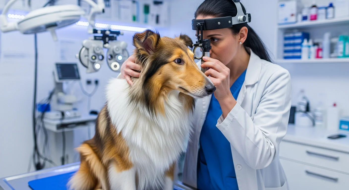
The primary instrument for fundus examination is the indirect ophthalmoscope. Unlike the direct ophthalmoscope, which most people have seen a doctor use — pressed close to the eye to view the retina — the indirect ophthalmoscope is worn on the examiner's head like a headlamp, and a handheld condensing lens is used to focus the image. This arrangement provides a wide-angle, stereoscopic view of the fundus with greater depth perception than the direct technique.
For CEA examination, the indirect ophthalmoscope is my primary tool. The wide field of view allows me to scan the entire fundus efficiently and to locate lesions that might be missed with the narrower view of direct examination. The stereoscopic vision provides crucial information about the three-dimensional structure of colobomas — the depth and contour of the excavation, not just its two-dimensional footprint.
For detailed examination of specific lesions, I may supplement with a slit lamp and contact lens, which provides magnified stereoscopic examination of the posterior segment. This combination — indirect for survey, slit lamp for detail — gives me the most complete picture of a dog's fundus.
Pupil Dilation: Essential for Complete Examination
Before fundus examination, I apply mydriatic drops — typically tropicamide — to dilate the pupils fully. An undilated pupil restricts the view of the fundus to a small central zone, potentially missing peripheral lesions entirely. CEA lesions are typically in the lateral (temporal) fundus, outside the region visible through an undilated pupil.
Pupil dilation takes approximately 20-30 minutes to reach maximum effect. The dilated state temporarily impairs the dog's ability to focus in bright light, though this causes no distress. Vision returns to normal within a few hours. I advise owners to allow for additional time at the clinic and to provide shade or a quiet environment for their dog while the dilation takes effect.
Dilation in Young Puppies
Very young puppies at the 6-8 week examination sometimes show less complete dilation than adults, partly because their pupillary light reflex is still maturing. This is usually not a significant obstacle for experienced examiners, but occasionally I will apply a second drop to optimise dilation before proceeding. The puppy screening examination protocol I use has been refined over many years to account for these practical realities of examining very young animals.
The Normal Canine Fundus
To recognise abnormality, one must first be thoroughly familiar with normality. The normal canine fundus has several characteristic features that vary with breed, coat colour, and pigmentation.
The tapetum is a reflective layer in the choroid that gives dogs their characteristic "eyeshine" in photographs taken with a flash. In dogs with normal tapetum, it appears as a brilliant reflective zone in the central and superior fundus, varying from blue-green to golden-yellow depending on the dog's age and pigmentation. The tapetum is absent in some dog breeds and in some individual dogs, resulting in a fully pigmented, non-reflective fundus — this is a normal variant, not a disease.
The non-tapetal fundus — the peripheral and inferior retina below and around the tapetum — is normally uniformly dark, pigmented by melanin in the retinal pigment epithelium. The choroidal blood vessels may or may not be visible through the pigment epithelium depending on the degree of pigmentation.
The optic disc is visible as a raised or flat oval structure, varying in appearance between breeds. In herding breeds, it tends to be oval with a slight horizontal orientation, and the myelin sheaths that surround optic nerve fibres as they emerge from the disc are often visible as pale extensions along the disc margins — an entirely normal finding in these breeds that should not be confused with pathology.
What CEA Lesions Look Like
When I examine a dog with choroidal hypoplasia, the abnormal area appears as a pale, usually mottled zone in the lateral fundus, typically outside the tapetal area. The normal dark uniformity of the non-tapetal fundus is interrupted by an area where the retinal pigment epithelium is thin and poorly pigmented, allowing the lighter scleral tissue to show through.
The choroidal blood vessels in this area may appear abnormal — irregular in pattern, reduced in density, or absent. The contrast between the normal dark non-tapetal fundus and the pale hypoplastic zone is usually clear, though it can be subtle in mildly affected animals or partially obscured by increasing pigmentation in older dogs.
The location is characteristically temporal to the optic disc — in the lateral fundus — though lesions can extend in various directions. Bilateral involvement with similar (though not always identical) findings in both eyes is the typical pattern, reflecting the bilateral nature of the developmental failure. I record the location, size (measured in disc diameters), and character of findings separately for each eye.
Understanding precisely what the choroidal hypoplasia grades represent in terms of what I observe allows breeders and owners to understand the significance of the numbers on their dog's examination report.
Recognising Colobomas
Colobomas are more dramatic ophthalmoscopic findings. Visible as excavations adjacent to the optic disc — most commonly extending from the disc margins into the surrounding tissue — they appear as darker, three-dimensionally deep defects where the normal tissue layers are absent. The stereoscopic view through the indirect ophthalmoscope allows me to appreciate the depth of the excavation, which is a key feature distinguishing a true coloboma from a flat area of tissue absence.
Around the coloboma margins, I often see areas of retinal disruption — irregular pigmentation, abnormal vessel patterns, and sometimes zones of retinal thinning that suggest the margins are under structural stress. These marginal changes concern me because they may indicate pre-detachment changes and can guide decisions about monitoring frequency or prophylactic intervention.
The size of colobomas relative to the optic disc diameter forms the basis of my grading. A coloboma that barely exceeds the disc in extent carries different implications from one that spans several disc diameters. My detailed description of CEA severity grades includes coloboma grading in clinical context.

The Importance of Specialist Examination
One of the most consistent findings in my career is that CEA diagnosis is significantly more accurate in the hands of specialist veterinary ophthalmologists than in general practice. This is not a criticism of general practitioners; fundus examination is a specialised skill that requires extensive training and ongoing experience to perform reliably.
Studies comparing general practice and specialist ophthalmology examination accuracy for CEA have shown meaningful differences in sensitivity and specificity. Mild choroidal hypoplasia in particular — the most common finding — can be subtle enough that inexperienced examiners may miss it or mischaracterise it. Conversely, normal variants such as myelin sheaths around the optic disc or irregular tapetal pigmentation can be misidentified as CEA by examiners not thoroughly familiar with the normal appearance of herding breed fundi.
For breeding certification purposes, I strongly recommend examination by a board-certified veterinary ophthalmologist (DACVO in North America, DECVO in Europe, RCVS specialist in the UK). The certification process that many breed clubs require specifically mandates specialist examination for this reason. The complete examination process including what happens before, during, and after the appointment, will help owners prepare for what to expect.
Recording and Reporting Findings
A thorough CEA examination produces a detailed written report that should include, for each eye:
- Pupil response and anterior segment findings
- Tapetal reflectivity and characteristics
- Specific description of any choroidal hypoplasia (location, size in disc diameters, character)
- Presence or absence of colobomas (location, size, depth assessment)
- Optic disc appearance
- Retinal vascular pattern and any areas of retinal change
- Overall clinical diagnosis and grade
This level of documentation is important for two reasons. First, it provides a baseline against which future examinations can be compared. Second, it creates a transparent record that breeders, breed clubs, and veterinary professionals can review independently. A report that simply states "CEA diagnosed" without specifics is inadequate for breeding programme purposes.
When I provide examination reports, I use standardised terminology and grade according to the same scale I have used throughout my career, ensuring consistency across different dogs and different time points. The combination of genetic testing with clinical examination records creates the most complete documentation possible for breeding decisions.
Retinal Photography and Imaging
Fundus photography — capturing photographs of the retina through specialist cameras — is available at some veterinary ophthalmology practices and provides permanent visual documentation of findings. Photographs allow direct comparison over time and can be shared with referring veterinarians and breed clubs as part of the health record.
Not all practices have fundus cameras, and the examination I perform ophthalmoscopically is not dependent on photography. But where it is available, I recommend fundus photography as a supplement to the written examination report for breeding dogs and for all dogs with documented colobomas. The visual record can be invaluable if disputes arise about a dog's health status or if comparison is needed between puppy and adult examinations.